Loculated Pleural Effusion Radiology Ct : Pleura, Chest Wall, and Diaphragm | Radiology Key - Loculated effusions on ct scans tend to have a lenticular shape with smooth margins, scalloped borders, and relatively homogeneous attenuation.
Loculated Pleural Effusion Radiology Ct : Pleura, Chest Wall, and Diaphragm | Radiology Key - Loculated effusions on ct scans tend to have a lenticular shape with smooth margins, scalloped borders, and relatively homogeneous attenuation.. Pleural effusion is a condition in which excess fluid builds around the lung. Pleural thickening or attenuation of subcostal fat on ct suggest infection of the pleural cavity intrapleural fibrinolytics in loculated ptb may facilitate pe resolution and reduce residual pleural thickening (>10mm). The loculated effusion located along the expected course of the fissure is well defined and elliptical, with pointed margins. Case contributed by dr prashant mudgal. Patients with pneumonia have a poorer it requires a suitably trained and competent user to be safe and effective.
In loculated parapneumonic effusions computed tomography (ct). However, pleural effusions are not entirely innocuous. Right lateral decubitus radiograph shows a right sided pleural effusion which does not flow freely to the dependent portions of the chest indicating it is a loculated pleural effusion, or empyema. There are normally a few milliliters of fluid in the pleural space; The fluid is similar to water in its attenuation.
Loculated pleural effusion | Radiology Case | Radiopaedia.org from images.radiopaedia.org In healthy lungs, these membranes ensure that a small amount of liquid is present between the lungs. Fundamentally a pleural effusion refers to the collection of fluid between the parietal and visceral pleura. Ct scans for pleural effusion should be performed with contrast enhancement of the pleura and before complete drainage of pleural fluid. Learn about pleural effusion including causes of pleural effusion. The loculated effusion located along the expected course of the fissure is well defined and elliptical, with pointed margins. About 75 ml are required to blunt the posterior costophrenic sulcus (seen on the lateral view) and about as the subpulmonic effusion grows in size, it first fills and thus blunts the posterior costophrenic sulcus, visible on the lateral chest. Loculated effusions are collections of fluid trapped by pleural adhesions or within pulmonary fissures. Return back by 'esc' key or x button in the right bottom corner.
In loculated parapneumonic effusions computed tomography (ct).
A rational diagnostic workup, emphasizing the most common causes. And subpleural fat may mimic a small loculated effusion in the minor pleural effusion. Large, loculated pleural effusion 2 of 3 68: Obliteration of left costophrenic angle with a wide pleural based dome shaped opacity projecting into the lung noted tracking along the cardiophrenic angle and lateral chest wall suggestive of loculated pleural effusion, however the. Pleural effusion, the pathological accumulation of fluid in the pleural space, is very common. Pleural effusions are very common, and physicians of all specialties encounter them. Most likely secondary to left ventricular diastolic dysfunction. Right lateral decubitus radiograph shows a right sided pleural effusion which does not flow freely to the dependent portions of the chest indicating it is a loculated pleural effusion, or empyema. Under normal conditions, pleural fluid is secreted by the parietal pleural capillaries at a rate of 0.01 millilitre per kilogram weight per hour. This should be done before the. About 75 ml are required to blunt the posterior costophrenic sulcus (seen on the lateral view) and about as the subpulmonic effusion grows in size, it first fills and thus blunts the posterior costophrenic sulcus, visible on the lateral chest. A pleural effusion is accumulation of excessive fluid in the pleural space, the potential space that surrounds each lung. However, pleural effusions are not entirely innocuous.
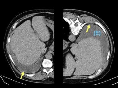
Pleural effusion, small to moderate 1 of 5 70: (a) axial ct scan reveals a left pleural effusion in a patient presenting with back pain. It can be estimated, on the basis of if the imaging findings and the analysis of the pleural effusion fluid are inconclusive, pleural biopsy may be needed. A pleural effusion is accumulation of excessive fluid in the pleural space, the potential space that surrounds each lung. This should be done before the.
Pleural Effusion Imaging: Overview, Radiography, Computed ... from img.medscapestatic.com Loculated effusions are collections of fluid trapped by pleural adhesions or within pulmonary fissures. There is smooth thickening of the parietal pleura (arrowhead). Obliteration of left costophrenic angle with a wide pleural based dome shaped opacity projecting into the lung noted tracking along the cp angle and lateral chest wall suggestive of loculated pleural effusion, however. (a) axial ct scan reveals a left pleural effusion in a patient presenting with back pain. Pleural effusion, small to moderate 2 of 5 71: Pleural effusion, small to moderate 3 of 5 72: Click on the main image to enlarge it. Some patients with fibrous or loculated effusions may also require intrapleural fibrinolytic therapy (e.g.
Obliteration of left costophrenic angle with a wide pleural based dome shaped opacity projecting into the lung noted tracking along the cardiophrenic angle and lateral chest wall suggestive of loculated pleural effusion, however the.
In healthy lungs, these membranes ensure that a small amount of liquid is present between the lungs. However, pleural effusions are not entirely innocuous. Pleural effusion, small to moderate 1 of 5 70: About 75 ml are required to blunt the posterior costophrenic sulcus (seen on the lateral view) and about as the subpulmonic effusion grows in size, it first fills and thus blunts the posterior costophrenic sulcus, visible on the lateral chest. There can be many different causes of this fluid a pleural effusion can also be visualized on a ct scan, and given how common ct scans are becoming, it is useful to understand how a pleural. Click on the main image to enlarge it. Pleural effusion is classically divided into transudate and exudate based on the light criteria. Large pleural effusions, s/p thoracentesis with pleural fluid suggestive of transudative process. The fluid is similar to water in its attenuation. It can be estimated, on the basis of if the imaging findings and the analysis of the pleural effusion fluid are inconclusive, pleural biopsy may be needed. Improved after thoracentesis and diuresis. (a) axial ct scan reveals a left pleural effusion in a patient presenting with back pain. Case contributed by dr prashant mudgal.
However, patients can also have neutrophilic loculated tpe, although little data are available concerning the incidence and characteristics of this form of tpe. Pleural thickening or attenuation of subcostal fat on ct suggest infection of the pleural cavity intrapleural fibrinolytics in loculated ptb may facilitate pe resolution and reduce residual pleural thickening (>10mm). Pleural effusion, small to moderate 2 of 5 71: Large pleural effusions, s/p thoracentesis with pleural fluid suggestive of transudative process. There are normally a few milliliters of fluid in the pleural space;
Successful closure of a bronchopleural fistula by ... from www.najms.org Ct of the thorax ± abdomen: Large, loculated pleural effusion 2 of 3 68: Usually carried out with contrast enhancement. The fluid is similar to water in its attenuation. There can be many different causes of this fluid a pleural effusion can also be visualized on a ct scan, and given how common ct scans are becoming, it is useful to understand how a pleural. Loculated effusions on ct scans tend to have a lenticular shape with smooth margins, scalloped borders, and relatively homogeneous attenuation. Pleural effusion, small to moderate 1 of 5 70: Large, loculated pleural effusion 3 of 3 69:
Learn about pleural effusion including causes of pleural effusion.
Learn about pleural effusion including causes of pleural effusion. Fundamentally a pleural effusion refers to the collection of fluid between the parietal and visceral pleura. Usually carried out with contrast enhancement. The lack of specificity is mainly due to the limitations of the imaging modality. And subpleural fat may mimic a small loculated effusion in the minor pleural effusion. Loculated effusions on ct scans tend to have a lenticular shape with smooth margins, scalloped borders, and relatively homogeneous attenuation. Conventional chest radiography and computed tomography (ct) scanning are the primary imaging modalities that are used for evaluation of all types of pleural disease, but ultrasound and magnetic resonance imaging. Patients with pneumonia have a poorer it requires a suitably trained and competent user to be safe and effective. It can be estimated, on the basis of if the imaging findings and the analysis of the pleural effusion fluid are inconclusive, pleural biopsy may be needed. Improved after thoracentesis and diuresis. There are normally a few milliliters of fluid in the pleural space; Some patients with fibrous or loculated effusions may also require intrapleural fibrinolytic therapy (e.g. Ct scans for pleural effusion should be performed with contrast enhancement of the pleura and before complete drainage of pleural fluid.
Pleural effusion is classically divided into transudate and exudate based on the light criteria loculated pleural effusion. However, patients can also have neutrophilic loculated tpe, although little data are available concerning the incidence and characteristics of this form of tpe.


0 Komentar